“Od 5 lat robimy to dobrze”… czyli rzecz o stymulacji Beta-CTF, krytycznej cząstki w chorobie Alzheimera
lipiec 24, 2015 by Jarek
Kategoria: Mózg, układ nerwowy
Ten wpis zaczynam od tłumaczenia maila, jaki przysłał mi Richard, gdy mu przesłałem najnowszy artykuł o chorobie Alzheimera: “To jest świetna informacja Jarek. Nie wiem czy pamiętasz ale około 5 lat temu omawialiśmy ten problem. Od 5 lat robimy to dobrze stosując DHA, zieloną herbatę i kurkumę, a teraz MgT. Robimy to dobrze!”
http://m.medicalxpress.com/news/2015-07-specific-protein-link-earliest-alzheimer.html
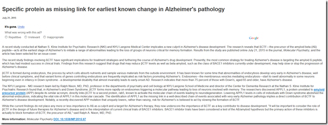
No to zaczynamy od początku. Właśnie pojawił się raport mówiący o istotnym czynniku inicjującym w bardzo wczesnym etapie, rozwój choroby Alzheimera. Mowa o Beta-CTF, proteinie-prekursorze amyloidu beta, która inicjuje utratę neuronów, krytycznych dla formowania się pamięci. Uważa się, że te badanie ma kluczowe znaczenie dla potencjalnych terapii, gdyż potwierdza, że leki które będą hamować aktywność genu BACE 1, z pewnością ograniczą rozwój choroby Alzheimera.
Co jest ważne, raport ten OPIERA SIĘ O BADANIA NAD OSOBAMI Z ZESPOŁEM DOWNA. W jego rozwinięciu jest przedstawiona następująca argumentacja, właśnie w oparciu o istniejącą sytuację z zespołu Downa:
1.BETA-CTF formuje się podczas endocytozy, procesu w którym następuje absorpcja cząsteczek odżywczych przez komórkę z poza niej. https://pl.wikipedia.org/wiki/Endocytoza
2.Nieprawidłowości w endocytozie są najwcześniejszym sygnałem rozwoju choroby Alzheimera, a poliformizmy genów odpowiedzialnych za ten proces są najczęściej najprecyzyjniejszym prognostykiem możliwości wystąpienia tej choroby.
3.Endosomy, które odpowiadają za sortowanie materiału pobranego na drodze endocytozy, w zespole Downa już od wieku niemowlęcego wykazują nieprawidłowości, co prowadzi do stanu wczesnego rozwoju choroby Alzheimera.
4.Badacze podczas tego projektu stwierdzili, że w ZD i w chorobie Alzheimera BETA-CTF tworzą się szybciej na endosomach, stąd bardzo szybka utrata nieodżywionych właściwie komórek a co za tym idzie pamięci.
5.Naukowcy odkryli cząsteczkę APPL 1 połączoną bezpośrednią z aktywnością BETA-CTF. Obniżając w ZD jej aktywność, likwiduje się złą endocytozę komórki.
Wrócę teraz do tego co omawialiśmy 5 lat temu z Richardem:
1.http://www.pnas.org/content/107/4/1630.long to był pierwszy artykuł, który opisywał w ZD problemy z BACE i BETA-CTF
2.Analizując literaturę uzyskaliśmy potwierdzenie, że DHA, EGCG, kurkuma ma działanie hamujące BACE i BETA-CTF.
http://www.ncbi.nlm.nih.gov/pubmed/15788759
3.A teraz jeszcze doszło MgT.
Co ja bym zrobił z moją pamięcią bez Richarda, nie wiem ale cieszę się że dzięki niemu już ponad 5 lat wyprzedzamy Świat w leczeniu naszych dzieci. Dzięki Richard!
Dodatkowa literatura:
Neuron. 2006 Jul 6;51(1):29-42.
Increased App expression in a mouse model of Down’s syndrome disrupts NGF transport and causes cholinergic neuron degeneration.
Salehi A, Delcroix JD, Belichenko PV, Zhan K, Wu C, Valletta JS, Takimoto-Kimura R, Kleschevnikov AM, Sambamurti K, Chung PP, Xia W, Villar A, Campbell WA, Kulnane LS, Nixon RA, Lamb BT, Epstein CJ, Stokin GB, Goldstein LS, Mobley WC.
Department of Neurology and Neurological Sciences, Stanford University, Stanford, California 94305, USA. asalehi@stanford.edu
Comment in:
· Neuron. 2006 Jul 6;51(1):1-3.
Degeneration of basal forebrain cholinergic neurons (BFCNs) contributes to cognitive dysfunction in Alzheimer’s disease (AD) and Down’s syndrome (DS). We used Ts65Dn and Ts1Cje mouse models of DS to show that the increased dose of the amyloid precursor protein gene, App, acts to markedly decrease NGF retrograde transport and cause degeneration of BFCNs. NGF transport was also decreased in mice expressing wild-type human APP or a familial AD-linked mutant APP; while significant, the decreases were less marked and there was no evident degeneration of BFCNs. Because of evidence suggesting that the NGF transport defect was intra-axonal, we explored within cholinergic axons the status of early endosomes (EEs). NGF-containing EEs were enlarged in Ts65Dn mice and their App content was increased. Our study thus provides evidence for a pathogenic mechanism for DS in which increased expression of App, in the context of trisomy, causes abnormal transport of NGF and cholinergic neurodegeneration.
PMID: 16815330 [PubMed – indexed for MEDLINE]
Proc Natl Acad Sci U S A. 2009 Dec 28. [Epub ahead of print]
Alzheimer’s-related endosome dysfunction in Down syndrome is A{beta}-independent but requires APP and is reversed by BACE-1 inhibition.
Jiang Y, Mullaney KA, Peterhoff CM, Che S, Schmidt SD, Boyer-Boiteau A, Ginsberg SD, Cataldo AM, Mathews PM, Nixon RA.
Center for Dementia Research, Nathan Kline Institute, Orangeburg, NY 10962 Departments of Psychiatry Physiology and Neuroscience Cell Biology, Langone School of Medicine, New York University, New York, NY 10016 Laboratory for Molecular Neuropathology, Mailman Research Center, McLean Hospital, Belmont, MA 02478 Department of Psychiatry and Neuropathology, Harvard Medical School, Boston, MA 02115.
An additional copy of the beta-amyloid precursor protein (APP) gene causes early-onset Alzheimer’s disease (AD) in trisomy 21 (DS). Endosome dysfunction develops very early in DS and AD and has been implicated in the mechanism of neurodegeneration. Here, we show that morphological and functional endocytic abnormalities in fibroblasts from individuals with DS are reversed by lowering the expression of APP or beta-APP-cleaving enzyme 1 (BACE-1) using short hairpin RNA constructs. By contrast, endosomal pathology can be induced in normal disomic (2N) fibroblasts by overexpressing APP or the C-terminal APP fragment generated by BACE-1 (betaCTF), all of which elevate the levels of betaCTFs. Expression of a mutant form of APP that cannot undergo beta-cleavage had no effect on endosomes. Pharmacological inhibition of APP gamma-secretase, which markedly reduced Abeta production but raised betaCTF levels, also induced AD-like endosome dysfunction in 2N fibroblasts and worsened this pathology in DS fibroblasts. These findings strongly implicate APP and the betaCTF of APP, and exclude Abeta and the alphaCTF, as the cause of endocytic pathway dysfunction in DS and AD, underscoring the potential multifaceted value of BACE-1 inhibition in AD therapeutics.
PMID: 20080541
APP in Pieces: βCTF implicated in Endosome Dysfunction
Research News in “The ALS Forum”, January 6, 2010
Among the products of amyloid precursor protein (APP) processing, the Aβ peptide claims the lion’s share of attention from researchers, but lately, other metabolites are grabbing the spotlight as potential purveyors of pathology. The APP intracellular domain (AICD), a product of γ-secretase cleavage and a putative transcriptional regulator, was recently implicated in neurodegeneration in mice (see ARF related news story on Ghosal et al., 2009). Now, a study from Ralph Nixon and colleagues at the Nathan Kline Institute, Orangeburg, New York, provides evidence that buildup of a different APP fragment, namely the C-terminal product of β-secretase cleavage (βCTF), causes the defects in endocytosis that are seen early in AD and can lead to neurodegeneration. The work suggests that inhibition of β-secretase may be the key to preventing the endosomal aspects of AD pathology.
With the field questioning whether Aβ is the full answer to AD, another metabolite of APP is of particular interest, especially given its pathogenic properties of disrupting crucial neuronal functions very early in AD, Nixon told ARF. The work appeared December 28 in the online edition of PNAS.
The study used fibroblasts from patients with Down syndrome (DS), who carry an extra dose of APP and are subject to early-onset AD. Like both AD and DS neurons, the fibroblasts show early endosome dysfunction characterized by increased endocytosis, enlargement of endosomes, and an increase in the number of the largest endosomes (Cataldo et al., 2008). Previous work from the lab of William Mobley at Stanford, in collaboration with Nixon’s lab, had shown that the disruption of endocytosis by APP overexpression in a mouse model of DS leads to neurodegeneration, as neurons are unable to support the retrograde transport of trophic factors necessary for their survival (see ARF related news story on Salehi et al., 2006). In the mouse neurons, the disruption of endocytosis depended on APP, but not necessarily on Aβ production.
In the new work, first author Ying Jiang shows that the endosome defects in DS fibroblasts depend on the overexpression of APP and production of the βCTF. RNAi knockdown (downregulation) of either APP or the β-secretase BACE1 restored both normal endosome function (measured by transferrin uptake) and morphology. Conversely, overexpressing APP or the βCTF in normal human fibroblasts induced endosome pathology. Jiang did not see the effect after transfecting a β-cleavage resistant APP mutant (M596V), establishing that β cleavage, and production of the βCTF, was critical to initiating the APP-induced endosome abnormalities.
To show that Aβ was not required, the researchers treated the fibroblasts with γ-secretase inhibitors, which decreased Aβ production while increasing levels of βCTF. In normal fibroblasts, the inhibitors caused an increase in lysosomal size, and in the DS fibroblasts, they worsened lysosomal pathology.
The results suggest that BACE inhibitors may have an advantage over Aβ-targeted therapies, at least where endosome dysfunction is concerned. I think that the BACE inhibitors are going to cover more bases in having a broader spectrum of effects on potential toxic metabolites of APP. I think our study gives more credibility to that approach for reversing endosome pathology, which we think is very critical to the development of the later pathologies, Nixon said.
How might βCTF affect endocytosis? In DS neurons, APP and βCTF are both elevated and distributed in early endosomes. We don’t know very much about what regulates endocytosis, but we strongly suspect that APP is involved, Nixon said. He cited work from Rachael Neve’s lab showing that APP binds to a protein complex that includes the GTP-binding protein Rab5 (Laifenfeld et al., 2007). Work that Nixon published a year ago with the late Anne Cataldo at McLean Hospital, Belmont, Massachusetts (see Cataldo et al., 2008), showed that Rab5 is markedly upregulated in the DS fibroblasts. When Anne manipulated Rab5 up or down, she essentially achieved the same circumstances that we achieved by manipulating βCTF, he said. Rab5 has become a focal point for us in initiating the downstream effects that lead to endocytosis upregulation and some of the adverse consequences of upregulation. We suspect that the βCTF is also involved as the mediating factor in the APP-endosome interaction that leads to Rab5 activation, or that it is involved in the signaling that is part of the Rab5 mechanism to stimulate endocytosis.
Neurons appear to be especially dependent on healthy endocytic function, Nixon said, and endosome dysfunction is increasingly related to a range of neurodegenerative diseases. A lot of genes, not only in AD, but in other neurodegenerative diseases, are starting to hit the endocytic pathway with a frequency that is much greater than expected by chance alone. In ALS, in Charcot-Marie-Tooth, and in some forms of spinal dementia, some genes that cause these conditions are very important functional proteins in either the early endosome or the late endosome. So there is a nexus of neurodegenerative phenomena associated with mutations anywhere along that endocytic pathway, which I think elevates the significance of endosomal dysfunction as a specific pathway to neurodegeneration in a number of diseases, he said.—Pat McCaffrey (Bio: see below).
Reference:
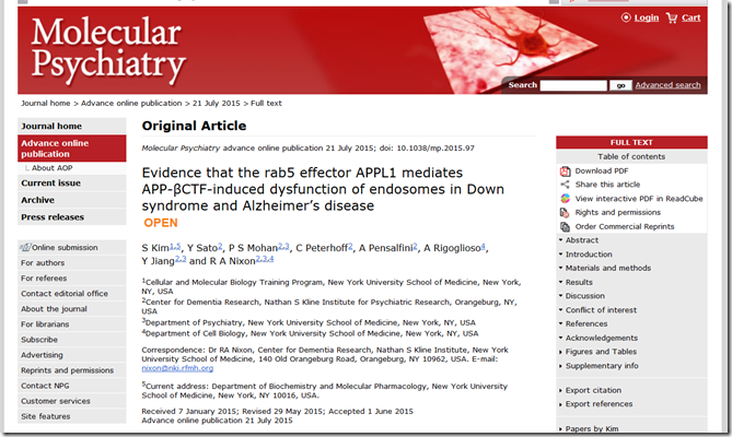
Jiang Y, Mullaney KA, Peterhoff CM, Che S, Schmidt SD, Boyer-Boiteau A, Ginsberg SD, Cataldo AM, Mathews PM, Nixon RA. Alzheimer’s-related endosome dysfunction in Down syndrome is Abeta-independent but requires APP and is reversed by BACE-1 inhibition. PNAS Early Edition, 2009 December 28.
http://www.researchals.org/page/4746/4632/0/2/
Pat McCaffrey, Science  Writer Pat McCaffrey is a freelance science writer specializing in biology and medicine. After earning an MS in Nutritional Biochemistry and a PhD in Applied Biology from MIT, Pat spent nearly 20 years doing research on cancer and immunology. Her research took her from Cambridge to Japan, where she lived and worked for two years. Then, it was back to Boston and positions at the Dana-Farber Cancer Institute and Vertex Pharmaceuticals. While at Vertex, Pat got a crash course in neurobiology when she was tapped to lead the company’s fledgling neurodegenerative disease program. Call it an early mid-life crisis, or simply the need to see her children before 7 pm every evening, but in 2001 Pat decided to abandon the life of a corporate scientist and pursue her childhood dream of being a writer. During an internship at the Harvard Medical School publications office, she met Tom Fagan, who introduced her to the Alzforum gang. So, for the next 20 years (at least), Pat plans to happily write about whatever topics come her way, with a special interest in oncology and neurological disease. In addition to writing for Alzforum, she freelances for Harvard Medical School and Yale Medical School. She is a regular contributor to the Lancet Oncology, and recently began to write for the Lancet Neurology journal.
Pat lives in Newton, MA, with her husband, a researcher at the Dana-Farber Cancer Institute. Above all, she treasures their summers in Rhode Island and any opportunity to wander the world with their three much-too-rapidly growing children.
http://www.alzforum.org/abo/arf/default.asp
Neuroreport. 2008 Aug 27;19(13):1329-33.
Epigallocatechin-3-gallate and curcumin suppress amyloid beta-induced beta-site APP cleaving enzyme-1 upregulation.
Shimmyo Y, Kihara T, Akaike A, Niidome T, Sugimoto H.
Department of Neuroscience for Drug Discovery, Graduate School of Pharmaceutical Sciences, Kyoto University, Kyoto, Japan. hsugimot@pharm.kyoto-u.ac.jp
Beta-site APP cleaving enzyme-1 (BACE-1), is a rate-limiting enzyme for beta amyloid production. Beta amyloid induces the production of radical oxygen species and neuronal injury. Oxidative stress plays a key role in various neurological diseases such as ischemia and Alzheimer’s disease. Recent studies suggest that oxidative stress induces BACE-1 protein upregulation in neuronal cells. Here, we demonstrate that naturally occurring compounds (-)-epigallocatechin-3-gallate and curcumin suppress beta amyloid-induced BACE-1 upregulation. Exposure of beta amyloid 1-42 to neuronal culture increased BACE-1 protein levels. (-)-Epigallocatechin-3-gallate or curcumin significantly attenuated beta amyloid-induced radical oxygen species production and beta-sheet structure formation. These two compounds have novel pharmacological effects that may be beneficial for Alzheimer’s disease treatment.
PMID: 18695518 [PubMed – indexed for MEDLINE]
Bioorg Med Chem Lett. 2003 Nov 17;13(22):3905-8.
Green tea catechins as a BACE1 (beta-secretase) inhibitor.
Jeon SY, Bae K, Seong YH, Song KS.
Division of Applied Biology & Chemistry, College of Agriculture & Life Sciences, Kyungpook National University, 1370 Sankyuk-Dong, Daegu 702-701, South Korea.
In the course of searching for BACE1 (beta-secretase) inhibitors from natural products, the ethyl acetate soluble fraction of green tea, which was suspected to be rich in catechin content, showed potent inhibitory activity. (-)-Epigallocatechin gallate, (-)-epicatechin gallate, and (-)-gallocatechin gallate were isolated with IC(50) values of 1.6 x 10(-6), 4.5 x 10(-6), and 1.8 x 10(-6) M, respectively. Seven additional authentic catechins were tested for a fundamental structure-activity relationship. (-)-Catechin gallate, (-)-gallocatechin, and (-)-epigallocatechin significantly inhibited BACE1 activity with IC(50) values of 6.0 x 10(-6), 2.5 x 10(-6), and 2.4 x 10(-6) M, respectively. However, (+)-catechin, (-)-catechin, (+)-epicatechin, and (-)-epicatechin exhibited about ten times less inhibitory activity. The stronger activity seemed to be related to the pyrogallol moiety on C-2 and/or C-3 of catechin skeleton, while the stereochemistry of C-2 andC-3 did not have an effect on the inhibitory activity. The active catechins inhibited BACE1 activity in a non-competitive manner with a substrate in Dixon plots.
PMID: 14592472 [PubMed – indexed for MEDLINE]


